You might think an MRI is just an MRI, but cardiac MRI is actually a whole different ball game. Let me explain what makes it special.
The Moving Target Problem
Your brain sits still. Your knee doesn’t wiggle. But your heart? It’s constantly beating, moving with every breath you take. That’s the core challenge—trying to get clear pictures of something that won’t hold still.
ECG Leads: Syncing with Your Heartbeat
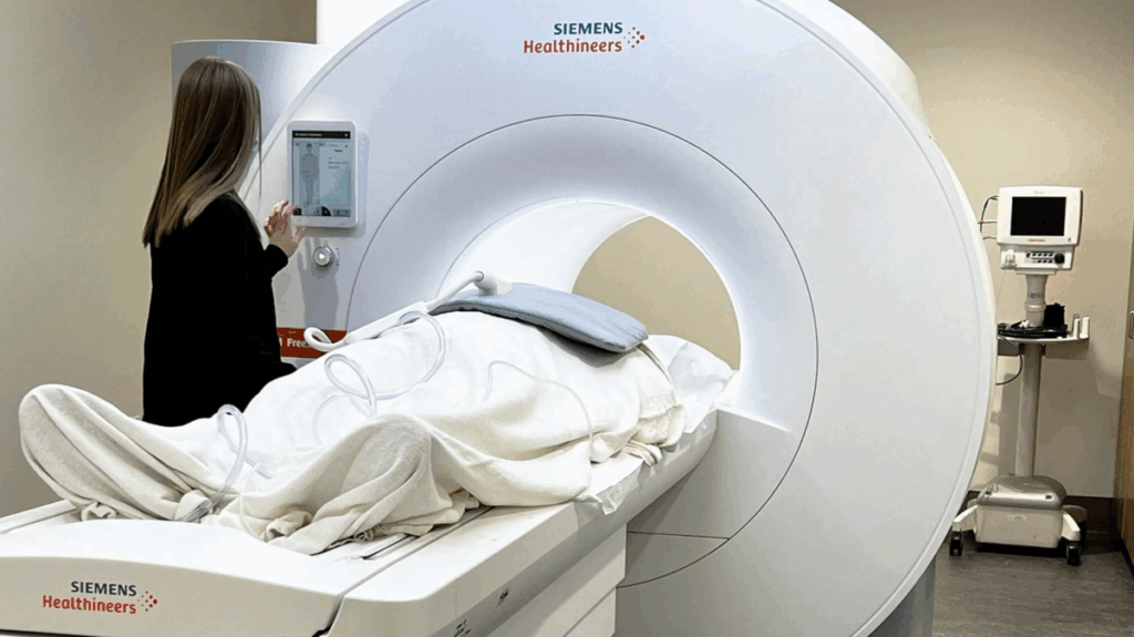
The biggest difference you’ll notice? They’ll attach ECG leads to your chest during a cardiac MRI, and the scanner uses the R wave from your ECG as a reference point to time when it captures data.
The scanner only grabs data during specific parts of your cardiac cycle, usually when your heart isn’t moving much. Images get created from data collected over multiple heartbeats, not just one. It’s like taking photos of a hummingbird only during the brief moments when it pauses mid-flight.
Regular MRIs don’t need any of this. They just scan continuously because whatever they’re imaging isn’t moving in a rhythmic pattern.
Breath-Holding
You’ll need to hold your breath multiple times during a cardiac MRI, usually for 7-15 seconds at a stretch. When you breathe, your heart shifts position in your chest, which would blur the images. Traditional MRIs sometimes ask you to stay still, but they’re nowhere near as strict about it.
What They’re Looking At
Cardiac MRI provides unique information. It can tell the difference between blood, muscle, clot, and scar tissue. It measures exactly how much blood your heart pumps with each beat and can spot damaged tissue that other tests miss.
Regular MRIs show you structure—what something looks like. Cardiac MRI shows you both structure and function—how your heart actually works. You get movie-like sequences showing your heart beating through its full cycle.
Time and Complexity
A cardiac MRI takes 30 minutes to 90 minutes. A regular MRI of your knee or shoulder? Usually 20-30 minutes. The extra time comes from all that synchronization with your heartbeat and multiple breath-holds.
Heart Rate Matters
Cardiac MRI works best when your heart rate is fairly regular. People with irregular rhythms can have trouble getting good images because the scanner gets confused about the timing. This isn’t an issue at all for traditional MRI—your heart rate could be all over the place and it wouldn’t affect a brain or spine scan.
Who Can’t Get One
Both types of MRI have restrictions about metal, but cardiac MRI has extra concerns. Many older pacemakers and defibrillators aren’t MRI-safe. Since these devices sit right near your heart in the area being scanned, it’s a bigger issue than with traditional MRI.
The Experience
Both are loud, but cardiac MRI is more interactive. The technologist will talk to you throughout, telling you when to breathe and when to hold your breath. Traditional MRI? You might just lie still for 20 minutes while they scan your shoulder.
FAQs
Do I need special preparation for cardiac MRI?
Not much different from regular MRI—no metal objects, tell them about implants. The main difference happens during the scan with the breath-holding and ECG leads. Some places might ask you to avoid caffeine beforehand.
Can I get a cardiac MRI with a pacemaker?
Depends on the model. Newer pacemakers are often MRI-conditional, meaning they can be scanned with special programming. Older ones usually can’t. Your cardiologist needs to coordinate with the MRI team.
Why does it take so much longer?
All that synchronization with your heartbeat adds up. The scanner waits for the right moment in your cardiac cycle over and over again through multiple beats. Plus multiple breath-holds and different types of scans being done in one session.
Is it better than an echocardiogram?
They’re different tools. Echo is quicker and cheaper. Cardiac MRI gives more precise measurements and better tissue information. Your doctor picks based on what they need to know.
Will I feel anything different?
You might feel the scanner timing with your heartbeat. The breath-holding can get tiring after doing it multiple times. If they’re doing a stress test, they might give you medication that makes your heart race temporarily.
Can it detect old heart attacks?
Yes, this is one of its strengths. It can show scar tissue from heart attacks that happened months or years ago. The pattern tells doctors which artery was blocked.
What if I can’t hold my breath that long?
Tell the technologist ahead of time. There are techniques that work with free breathing, though image quality might not be quite as good. If you have severe lung disease, let your doctor know—they might need alternative imaging.
Does insurance cover it?
Usually covered when medically necessary, but might need pre-authorization. It’s typically more expensive than regular MRI because of the complexity involved.
Takeaway
Cardiac MRI might look like any other MRI scan from the outside, but what happens inside that machine is far more sophisticated. All that heartbeat synchronization, breath-holding, and functional imaging adds up to something pretty remarkable—detailed pictures and measurements that can spot heart problems other tests might miss.
If your doctor orders one, now you know why it takes longer and what all that extra effort is actually accomplishing. Your heart’s worth the extra time.